Hosted by
The Espians
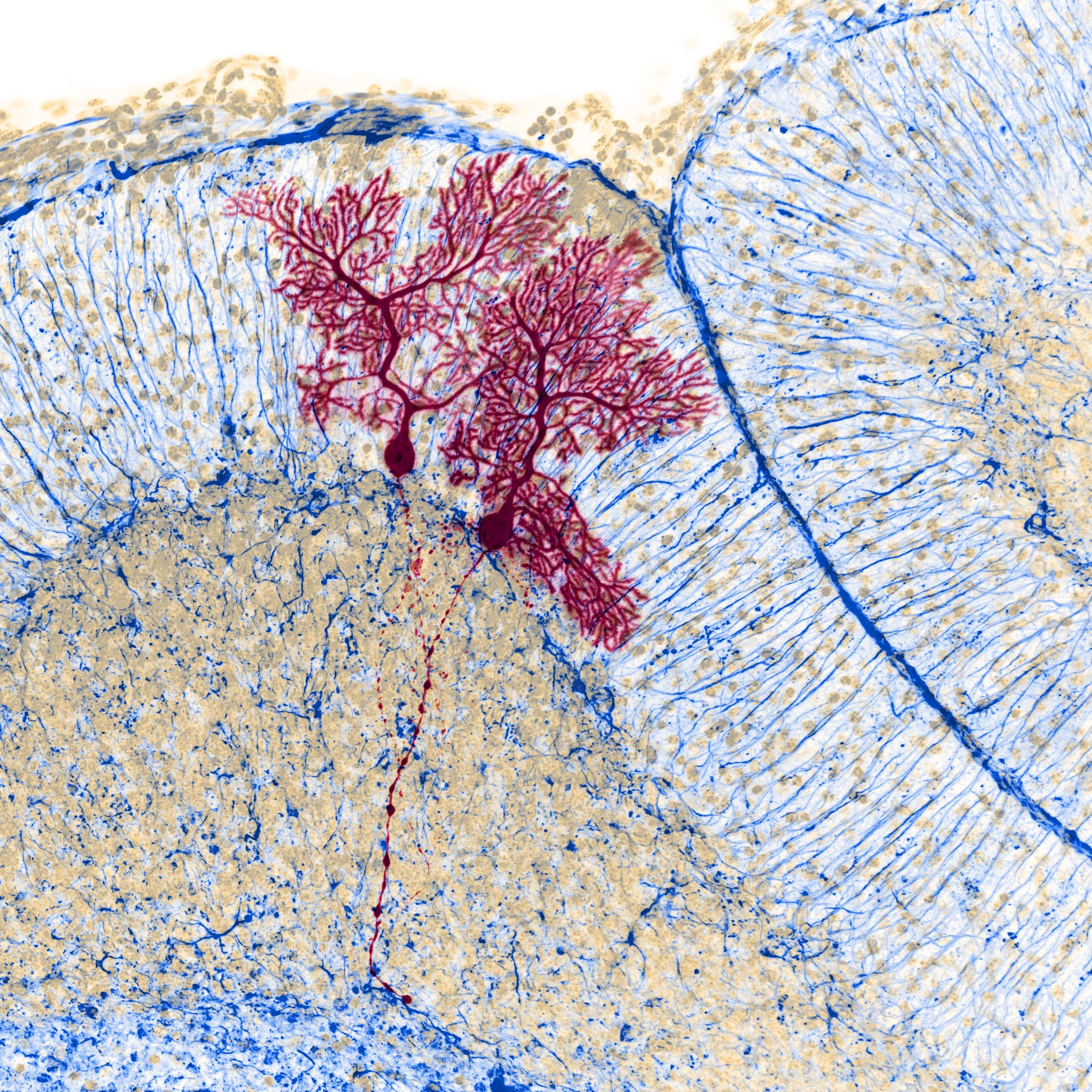
[Permanent link] Fixed mouse cerebellar brain section, 300 μm
Zoom: 1× 2× Fit monitor
Remove cookie
To enable HTML5 features, use a modern, JavaScript-enabled browser such as Google Chrome or Mozilla Firefox!
Faint-orange: DAPI; Red: biocytin filled Purkinje cells visualized by streptavidin 488; Blue: GFAP labeled with anti-GFAP, visualized by CF568 secondary antibody.
Confocal, 20×
Department of Molecular and Developmental Neurobiology, Institute of Experimental Medicine (KOKI), Hungarian Academy of Sciences (MTA)
Benjamin Barti
2018 Nikon-KOKI image contest, Nikon Center of Excellence
