Legend
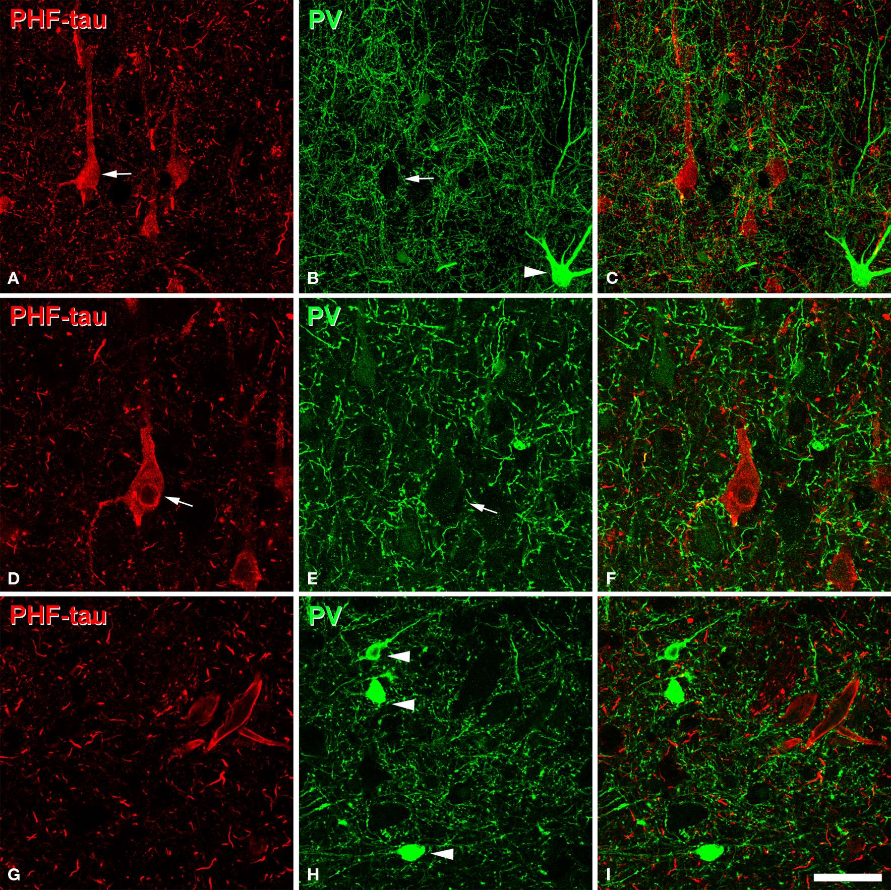
(A,B), pairs of confocal images (stacks of 10 optical sections; step size: 1. 04 μm) to illustrate that PV-ir axon terminals (green) are preserved around the somata of PHF-tau-ir neurons (red) in CA1 (patient P7). (D,E), pairs of single confocal sections showing an PHF-tau-ir pyramidal cell (also indicated in (A) with an arrow) to illustrate its perisomatic innervation by PV-ir axon terminals at higher magnification. (G,H), pairs of single confocal sections showing three PV-ir neurons (arrowhead), none of them are labeled for PHF-tau. Panels (C), (F), and (I) were obtained after combining images (A) and (B), (D) and (E), and (G) and (H), respectively. Scale bar: (A.C), 67 μm; (D.I), 43 μm.
Technique
Confocal
Citation
Pericellular innervation of neurons expressing abnormally hyperphosphorylated tau in the hippocampal formation of Alzheimer's disease patients, Lidia Blazquez-Llorca et al., Neuroanatomy 10.3389/fnana.2010.00020 (2010)
