Legend
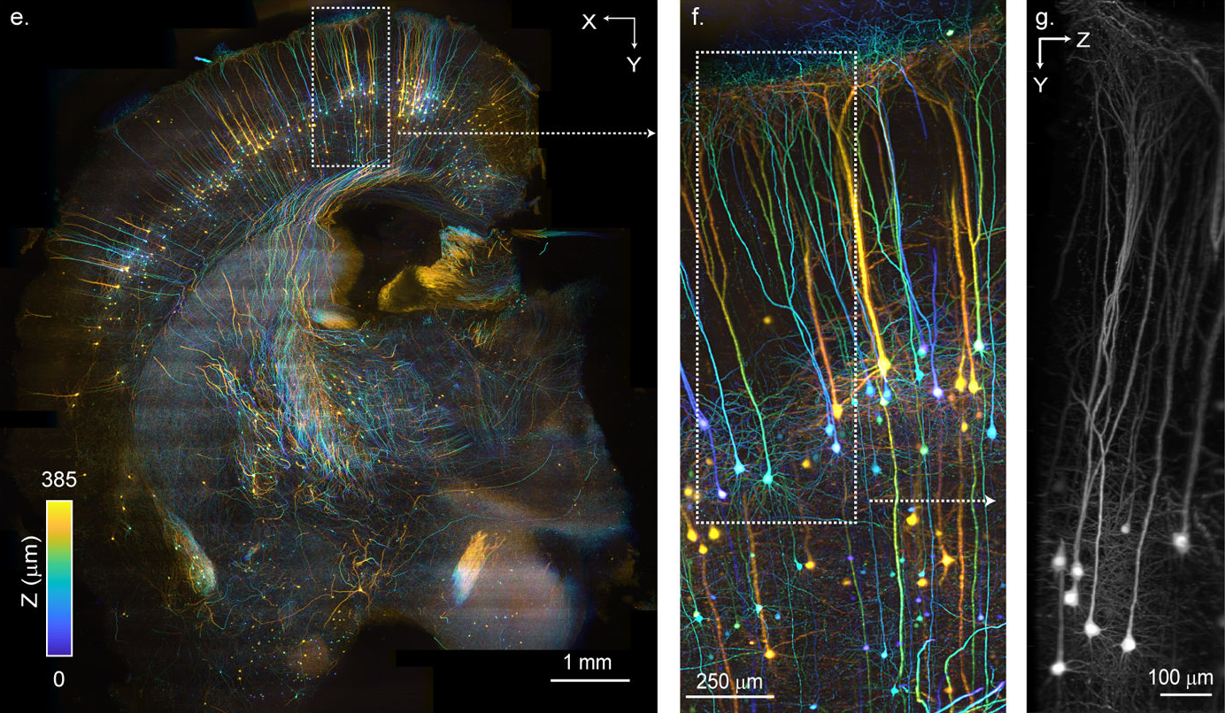
The authors demonstrate a new microscopy technique called SCAPE 2.0 (Swept, Confocally Aligned Planar Excitation) that can achieve high-resolution volumetric imaging at a high enough framerate to enable realtime imaging. The figures show the system imaging an mCUBIC6, cleared Thy1-GFP mouse brain that was sliced coronally and imaged from below in a glass-bottomed Petri dish. 18 adjacent, overlapping volume strips, 1.10 mm wide in y and 456 microns deep were acquired with 1 micron x-spacing at 520 fps and stitched and cropped into a ~8.37 × 9.06 × 0.385 mm volume with a sampling density of 1 × 1.37 × 1.14 μm x-y-z. Total acquisition took 242 seconds. Color depth-encoding demonstrates how SCAPE 2.0’s near-isotropic sampling enables clear tracing of axons and dendrites across large areas of the intact brain.
(e) Color-depth encoded top-down view of a 385 µm thick volume acquired on a coronal hemi-section of an mCUBIC-cleared Thy1-GFP brain using stage-scanning and stitching. Scale bar: 1 mm. (f) Zoomed view of subregion outlined in (e). (g) YZ plane maximum intensity projection, taken along the X direction of the subregion outlined in (f) demonstrating SCAPE 2.0’s near isotropic sampling and resolution
Press release
Technique
SCAPE 2.0
Location
USA, Japan
Citation
Real-time volumetric microscopy of in-vivo dynamics and large-scale samples with SCAPE 2.0. Venkatakaushik Voleti, Kripa B. Patel, Wenze Li, Citlali Perez Campos, Srinidhi Bharadwaj, Hang Yu, Caitlin Ford, Malte J. Casper, Richard Wenwei Yan, Wenxuan Liang, Chentao Wen, Koutarou D. Kimura, Kimara L. Targoff, and Elizabeth M.C. Hillman. Nature Methods. 2019 Oct; 16(10): 1054–1062. doi: 10.1038/s41592-019-0579-4
