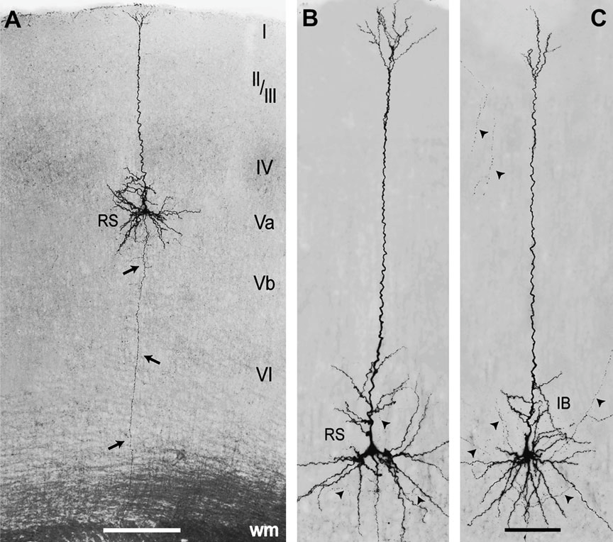
Extended-focus views of layer Va pyramidal cells stained for biocytin. (A) Pyramidal cell showing an RS firing pattern located below the lateral aspect of a layer IV barrel with its apical dendrite reaching the pial surface and the axon (arrows) reaching the white matter (wm). (B, C) Higher magni.cations of an RS (B) and an IB pyramidal cell (C) with some captured axon fragments (arrowheads). Note that no obvious qualitative differences are visible between the IB and RS neuron. Scale bars: 250 μm(A); 50 μm(B, C).
“Morphology, electrophysiology and functional input connectivity of pyramidal neurons characterizes a genuine layer va in the primary somatosensory cortex”, D. Schubert, R. Kötter, H.J. Luhmann and J.F. Staiger, Cerebral Cortex 2006 Feb, 16(2):223–36
